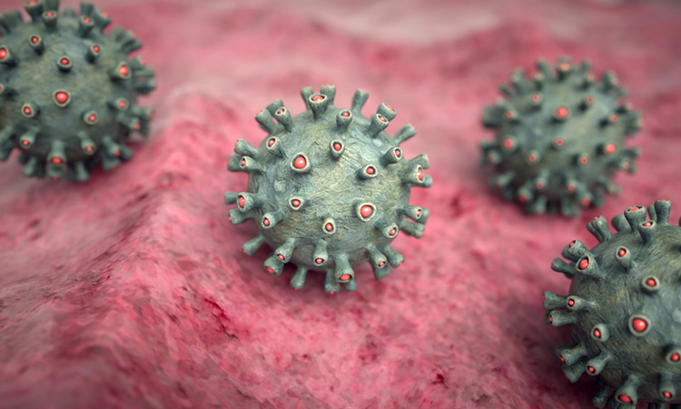
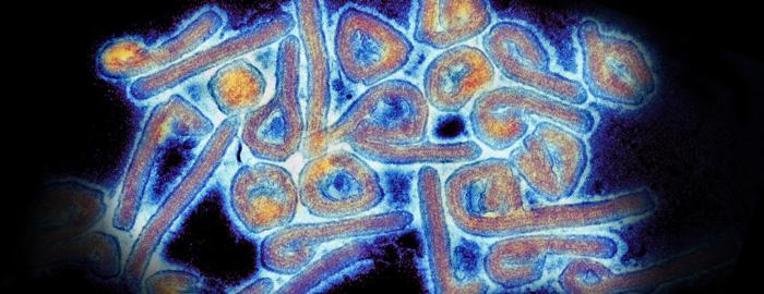
Marburg Virus Infected Cells
Marburg virus in the liver of an experimentally infected monkey. Lab workers got infected.
Antibodies And Remdesivir Used To Treat Marburg Virus In Rhesus Macaques
In addition those MARV-infected cells con-tained VP30-GFPlabeled NP-positive filamentous structures which had an average length of 962 100 nm n 50 Fig.

Marburg virus infected cells. It was discovered that year during a set of outbreaks of Marburg virus disease in the German cities of Marburg and Frankfurt and the Yugoslav capital BelgradeLaboratory workers were exposed to tissues of infected grivet monkeys the African green monkey Chlorocebus aethiops at the Behringwerke a major industrial plant in Marburg which was. The Marburg virus can also be passed indirectly from person to person through contaminated objects or materials containing infected body fluids. African fruit bats Rousettus aegyptiacus flying outside a cave and observation platform in western Uganda.
However for the 2 cases in tourists visiting Uganda in 2008 unprotected contact with infected bat feces or aerosols are the most likely routes of infection. This infection is extremely destructiveshortly after this phase of infection the liver cells are destroyed. In general Marburg virus entry into infected cells consists of attachment endocytosis and fusion.
For the Marburg and Ebola viruses related viruses that cause severe hemorrhagic fevers the mechanism of nucleocapsid transport remains poorly understood. When the virus interacts with Keap1 Marburg virus-infected cells survive longer facilitating virus growth. This protein mediates infection by binding to the viral envelope glycoprotein.
This can include medical equipment like needles or cell cultures as well as more common objects like soiled bed linens. Studying Ebola virus they. We have applied cryo-electron tomography and sub-tomogram averaging methods to derive structures of Marburg virus a highly pathogenic filovirus both after release and during assembly within infected cells.
The most dangerous viruses of all time. At the specified time points after infection cell supernatants were harvested and evaluated for virus load. When we generated 3-D images of virus particles frozen in the act of assembling and budding from infected cells however we found that despite their structural similarities Marburg virus particles are released from infected cells with the pointed end of the nucleocapsid facing out whereas rabies virus is released with its barbed end facing out suggesting different budding mechanisms.
Live-cell imaging of Marburg virus-infected cells uncovers actin-dependent transport of nucleocapsids over long distances. Live-cell imaging of Marburg virus-infected cells uncovers actin-dependent transport of nucleocapsids over long distances Gordian Schudt Larissa Kolesnikova Olga Dolnik Beate Sodeik and Stephan Becker. The research builds on previous research in Dr.
The reservoir host of Marburg virus is the African fruit bat Rousettus aegyptiacus. Huh-7 cells were infected with rMARV wt or rMARV PSAPmut. At 28 h pi cells were processed in two ways i fixed scraped pelleted and then embedded in Epoxy resin A and C.
Primates including humans can become infected with Marburg virus and may develop serious disease with high mortality. Inclusions in rMARV PSAPmut infected cells are more densely packed with nucleocapsids than inclusions in rMARV wt infected cells. Or ii fixed and embedded in Epoxy resin on Thermanox slides B and D.
Gordian Schudt Institut für Virologie Philipps-Universität Marburg D-35043 Marburg Germany. Both attachment and fusion are mediated by Marburg virus glycoprotein where it binds to. Virions bud off the surface membrane of liver cells and accumulate in the narrow spaces between cells.
Marburg virus was first described in 1967. Marburg was first noticed in 1967 after outbreaks in Germany and Serbia. For Marburg and Ebola that element is the NiemannPick C1 NPC1 membrane protein 1.
It is unknown how Marburg virus first transmits from its animal host to humans. VP30-GFP was colocalized with inclusions in MARV-infected cells Fig. Growth of EBOV and MARV in DCs A or Vero E6 cells B.
Here we developed and used live-cell imaging of fluorescently labeled viral and host proteins to characterize the dynamics and molecular requirements of nucleocapsid transport in Marburg virus-infected cells under biosafety level 4. DCs or Vero E6 cells were infected with EBOV or MARV at an MOI of 1. Fruit bats infected with Marburg virus do not to show obvious signs of illness.
Growth and visualization of Ebola virus EBOV and Marburg virus MARV in human dendritic cells DCs. For the Marburg Virus to infect the hosts cell an essential element is needed.
The Next Pandemic Marburg Gavi The Vaccine Alliance

First West African Case Of Marburg Virus Detected Abs Cbn News
Marburg Virus Disease Causes Symptoms Diagnosis Treatment Prevention

Marburg Virus Structure And Transmission

A Survival Curve Of Guinea Pigs Gps Infected With Marburg Virus Ravn Download Scientific Diagram
Marburg Virus Survivor Holds Treatment Clues

Experimental Drug Stops Ebola Like Infection Science Aaas
/Marburgvirus-6b3a8045821e4e3a8a919e8231033555.jpg)
Marburg Virus Symptoms Causes Diagnosis And Treatment
Marburg Virus What To Know About Di Virus Wey Dem Confam For Guinea West Africa Bbc News Pidgin
Posting Komentar untuk "Marburg Virus Infected Cells"