Marburg Virus Genome Structure
The two species Marburg and Ebola virus are genetically distinct with 7 genes and a total molecular length of approximately 19 kb 19112 bp making them the owners of the largest known genomes of negative-strand RNA viruses. There are two peaks of nucleotide coverage in that genome situated in the part 2 of the GP coding region and in part 1 of the VP30 coding region.
It has been causing sporadic outbreaks in Central Africa since at least 1975 2.
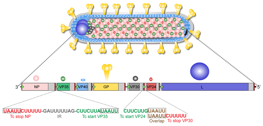
Marburg virus genome structure. Marburg virus MARV encodes a nucleoprotein NP to encapsidate its genome by oligomerization and form a ribonucleoprotein complex RNP. Request a Free Quote Today. Genomic RNAs are not polyadenylated at their 3ends and there is no evidence of 5-end terminal cap structures or covalently linked proteins.
Marburg virus has an unusual shape. Marburg virus is a hemorrhagic fever virus of the Filoviridae family of viruses and a member of the species Marburg marburgvirus genus MarburgvirusMarburg virus MARV causes Marburg virus disease in humans and other primates a form of viral hemorrhagic fever. If Marburg virus orients its RNA as RSV does the three-prime end of its genome would also be the first to bud as it is in VSV.
These genomes are 191 kb. The viral RNA dependent RNA polymerase binds the encapsidated genome at the leader region then sequentially transcribes each genes by recognizing start and stop signals flanking viral genes. The virus is considered to be extremely dangerous.
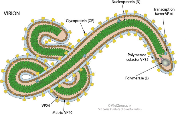
Marburg virions the entire virus particle may be coiled or branched but are typically 80nm in width and vary in length 795-828nm 2. Sequence analysis of the second through the sixth genes of the Ebola virus EBO genome indicates that it is organized similarly to rhabdoviruses and paramyxoviruses and is virtually the same as Marburg virus MBG. RNA editing may occur at NP gene.
These types of viruses encode their genome in the form of single stranded negative polarity RNA. Request a Free Quote Today. The virus RdRp partially uncoats the nucleocapsid and transcribes the genes into positive-stranded mRNAs which are then translated into structural and nonstructural proteins.
Similar situation can be found in the Marburg virus genome Table 3. Both of them demonstrate extremely low. Marburg virus MARV belongs to the family Filoviridae as a member of the species Marburg marburgvirus 1.
The virus consists of a nucleocapsid surrounded by a cross-striated helical capsid. Similar to EBOV it causes severe haemorrhagic fever in humans and. They are pleomorphic in shape which means they can be a number of different shapes are rod-like or ring-like crook- or six-shaped or with branched structures.
The gene order - 3 untranslated region-NP-VP35-VP40-GP-VP30-VP24-L-5 untranslated region-resembles that of other non-segmented negative-strand NNS RNA viruses. According to previous investigation on nonsegmented negative-sense RNA viruses nsNSV the newly synthesized NPs must be. In that virus the earliest synthesized part of the genome the so-called three-prime end is packed in the barbed end while in a related virus respiratory syncytial virus RSV it is in the pointed end.
The genome of Marburg virus MBG a filovirus is 191 kb in length and thus the largest one found with negative-strand RNA viruses. 42 Marburg virus MARV a close relative of the Ebola virus EBOV belongs to the 43 family Filoviridae and possesses a 191 kb non-segmented single-stranded negative-sense 44 RNA genome vRNA1. Experimental DesignLibrary PreparationSequencingBioinformatic Analysis.
Experimental DesignLibrary PreparationSequencingBioinformatic Analysis. The Marburg virus is part of the filovirus family 1. Marburgvirus genomes are linear non-segmented RNA molecules of negative polarity.
MRNAs are capped and polyadenylated by the L protein during synthesis. Their basic structure is long and filamentious essentially bacilliform but the viruses often takes on a U shape and the particles can be up to 14000 nm in length and average 80 nm in diameter. The World Health Organization WHO rates it as a Risk Group 4 Pathogen.
Marburgvirus L binds to a single promoter located at the 3 end of the genome.

Ebolavirus An Overview Sciencedirect Topics

Filovirus An Overview Sciencedirect Topics
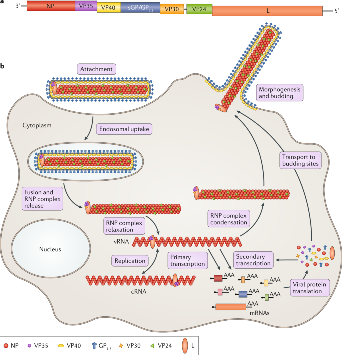
Therapeutic Strategies To Target The Ebola Virus Life Cycle Nature Reviews Microbiology

The Genome And Virus Structure Of The Filoviridae A Linear Download Scientific Diagram
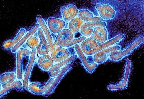
Marburg Virus Structure And Transmission

Viruses Free Full Text Forty Five Years Of Marburg Virus Research Html

Rodent Adapted Filoviruses And The Molecular Basis Of Pathogenesis Abstract Europe Pmc

The Genome And Virus Structure Of The Filoviridae A Linear Download Scientific Diagram
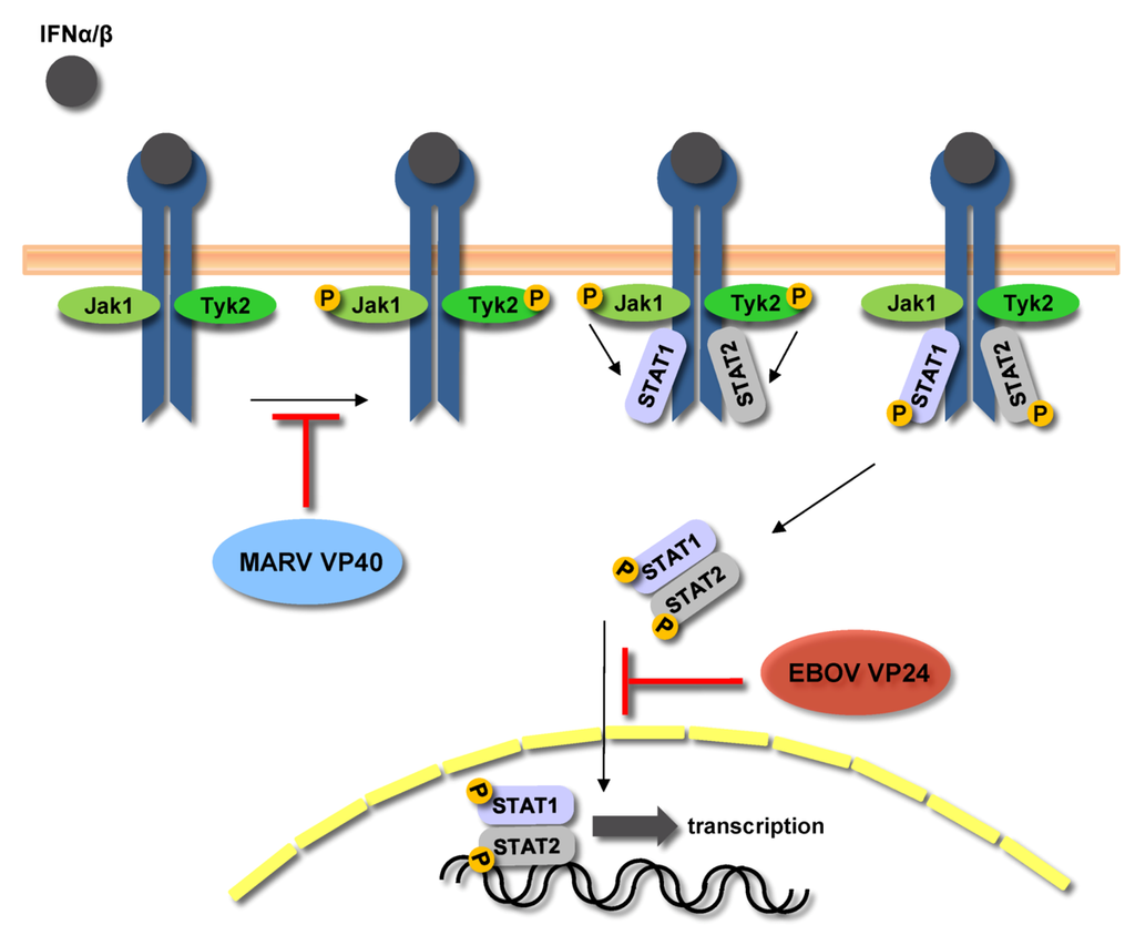
Viruses Free Full Text Forty Five Years Of Marburg Virus Research Html

Posting Komentar untuk "Marburg Virus Genome Structure"