Marburg Virus Replication
We show here that Marburg virus replicates in endothelial cells almost as well as in monkey kidney cells commonly used for virus propagation. Six species of polyadenylated subgenomic RNAs.
Research Muhlberger Lab
Marburg virus a filovirus.
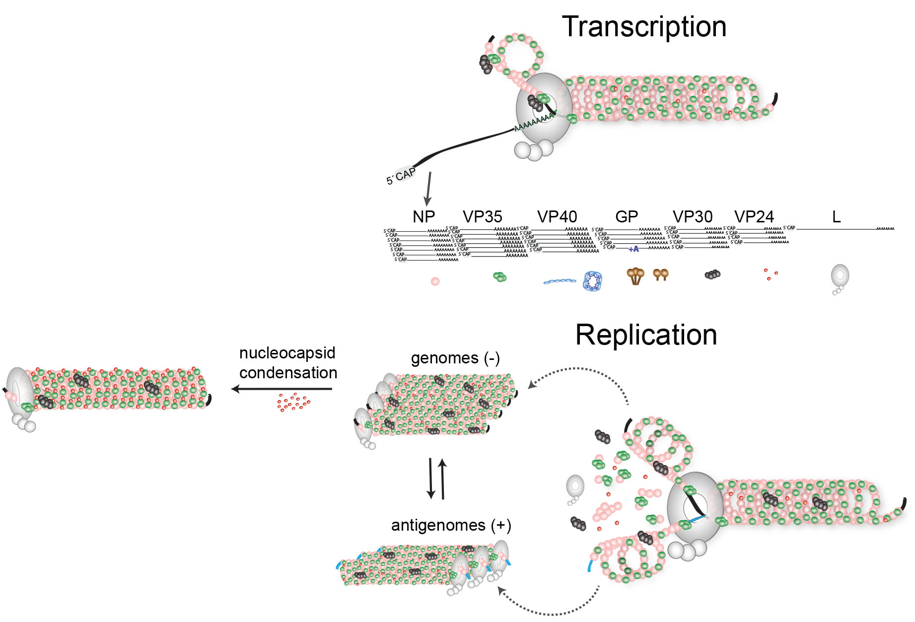
Marburg virus replication. The gene order - 3 untranslated region-NP-VP35-VP40-GP-VP30-VP24-L-5 untranslated region-resembles that of other non-segmented negative-strand NNS RNA viruses. Ribonucleocapsid is released into the cytoplasm. Marburg and Ebola viruses cause similar pathological changes in humans.
Replication presumably starts when enough nucleoprotein is present to encapsidate neo-synthetized antigenomes and genomes. Three of the four nucleocapsid proteins of Marburg virus NP VP35 and L are sufficient to mediate replication and transcription of Marburg virus-specific monocistronic minigenomes. Three of the four nucleocapsid proteins of Marburg virus NP VP35 and L are sufficient to mediate replication and transcription of Marburg virus-specific monocistronic minigenomes.
The most striking lesions are found in the liver spleen and lymph nodes. In women who have been infected while pregnant the virus persists in the placenta amniotic fluid. Méssenger RNAs gene order and regulatory elements of the replication cycle Anthony Sanchez Received 22 October 1991revision received 12 December 1991accepted 13 December 1991 SummaryThe genome of Marburg virus MBG a filovirus is 191 kb in length and thus the largest one found with negative-strand RNA viruses.
Fusion of virus membrane with the vesicle membrane. These sites include the testicles and the inside of the eye. Macrophages monocytes and dendritic cells are the primary sites of infection where the virus replicates to high titer.
Our data support the concept that the destruction of endothelial cells resulting from Marburg virus replication is a possible mechanism responsible for the hemorrhagic disease and the shock syndrome typical of this infection. The genome of Marburg virus MBG a filovirus is 191 kb in length and thus the largest one found with negative-strand RNA viruses. Mühlberger E Lotfering B Klenk H-D Becker S.
Unlike Ebola virus a different host protein was shown to interact with Marburg virus. Replication and transcription of the viral genome takes place in the cytoplasm of the host and the virions exit the host by a process known as budding a process during which the virus steals some of the cells membrane 1. Marburg virus enters cells by endocytosis and fusion meaning the virus is engulfed by the host and the viral membrane fuses with the host membrane.
Sequential transcription viral mRNAs are capped and polyadenylated by polymerase stuttering in the cytoplasm. J Virol 1998 72. If we can develop inhibitors the virus will die and replicate more slowly -- thats the hypothesis that we.
Persistent virus in people recovering from Marburg virus disease Marburg virus is known to persist in immune-privileged sites in some people who have recovered from Marburg virus disease.

Pin On Virology
Viruses Free Full Text Forty Five Years Of Marburg Virus Research Html

Filoviridae Sciencedirect

Figure 1 From Filovirus Pathogenesis And Immune Evasion Insights From Ebola Virus And Marburg Virus Semantic Scholar

Marburg Hemorrhagic Fever Pathogenesis Model Download Scientific Diagram

Filovirus Pathogenesis And Immune Evasion Insights From Ebola Virus And Marburg Virus Abstract Europe Pmc

Filovirus An Overview Sciencedirect Topics

The Typical Different Stages Of Virus Life Cycle 1 Attachment In Download Scientific Diagram

Figure 4 From Filovirus Pathogenesis And Immune Evasion Insights From Ebola Virus And Marburg Virus Semantic Scholar
Viruses Free Full Text Forty Five Years Of Marburg Virus Research Html
Posting Komentar untuk "Marburg Virus Replication"